Konica Minolta Healthcare Americas, Inc. is pleased to announce the publication of a study that found Dynamic Digital Radiography of the chest delivers similar efficacy as lung ventilation perfusion for the detection of chronic thromboembolic pulmonary hypertension.
Published online November 8 in the journal, Radiology, the study enabled the visualization of pulmonary hemodynamics and perfusion with high sensitivity, specificity and diagnostic accuracy, and without the use of contrast media.
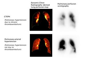
WAYNE, N.J., Nov. 22, 2022 (GLOBE NEWSWIRE) -- Konica Minolta Healthcare Americas, Inc., a leader in medical diagnostic imaging and healthcare information technology, is pleased to announce the publication of a study that found Dynamic Digital Radiography (DDR) of the chest (aka DCR) delivers similar efficacy as lung ventilation perfusion (V/Q) for the detection of chronic thromboembolic pulmonary hypertension (CTEPH). In addition, DDR was performed without the need for contrast media and at a significantly lower radiation dose to the patient, approximately one-tenth of lung V/Q scintigraphy. The study by lead author Yuzo Yamasaki, MD, PhD, from Kyushu University (Fukuoka, Japan), was published November 8 in Radiology, the scientific journal of the Radiological Society of North America.
Dynamic Digital Radiography is a radiographic technique that provides a series of individual digital X-ray images acquired at high speed and low radiation dose. These images provide a diagnostic-quality view of anatomical structures in motion and the ability to visualize the dynamic interaction with physiological changes over time. DDR provides enriched radiographic information and enhanced diagnostic capabilities such as the ability to visualize pulmonary hemodynamics without contrast media, which is the subject of this research. CTEPH is a complication of pulmonary embolism and a significant cause of pulmonary hypertension with associated morbidity and mortality if left untreated. As a potentially treatable cause of pulmonary hypertension, identification of CTEPH with imaging may significantly impact clinical outcomes of patients with this condition.
In this retrospective study, 50 patients with pulmonary hypertension diagnosed via right heart catheterization and invasive pulmonary angiography underwent dynamic X-ray imaging, with 7-10 seconds of inspiratory breath holding, using a Konica Minolta AeroDR® HD flat panel detector and pulsed X-ray system (RADspeed Pro, Shimadzu). Each patient also had conventional chest radiography and underwent a lung V/Q study on a SPECT/CT system with intravenous injection of technetium-99m macroaggregated albumin, 185 MBq. Of the 50 patients, 29 met the World Health Organization (WHO) criteria for CTEPH and 21 were classified as non-CTEPH.
Dr. Yamasaki and co-authors reported similar sensitivity and diagnostic accuracy of DDR (97% and 92%, respectively) compared to V/Q (100% and 94%, respectively) with the same specificity (86%). The agreement between DDR and V/Q interpretations was substantial (κ = 0.79 [95% CI: 0.61, 0.96] percent agreement=0.9 [95% CI: 0.79, 0.95]) and the interobserver agreements for both DDR and V/Q scans (κ = 0.71 and κ = 0.73, respectively) were also significant. Two chest radiologists evaluated the DDR images and two nuclear medicine physicians evaluated the V/Q data.
The authors note that while early diagnosis of CTEPH is important for better patient outcomes, V/Q scanning is underused and there is a clinical need for an easy, noninvasive screening method. Compared to other modalities, including dual-energy CT and MRI, the authors wrote that DDR has several advantages, including low radiation dose and no contrast media or radionuclides. Additionally, both dual-energy CT and MRI are more technically challenging, more expensive, have limited availability, and lack multicenter validation. The authors conclude that DDR “may be a low-cost alternative to V/Q scanning for the diagnosis of this disease,” and they further note that the DDR system can also be used as a conventional X-ray system and installed in a smaller space than what is needed for a SPECT/CT system.
In an accompanying editorial, John C. Wandtke, MD, professor, and Katherine Kaproth-Joslin, MD, PhD, Department of Imaging Sciences, University of Rochester Medical Center, comment that a major advantage is that the radiation dose is approximately one-tenth that of lung V/Q scintigraphy, one-twentieth that of standard CT pulmonary angiography, and one-fifth that of low-dose CT pulmonary angiography. The authors further highlight the clinical value of DDR for CTEPH. They wrote that DDR may be a reliable alternative to contrast- and radionuclide-enhanced imaging methods and it “appears to represent an innovative method for fast and cost-effective screening of patients for CTEPH.”
“The ability to visualize pulmonary hemodynamics without contrast using images acquired with standard X-ray systems is groundbreaking. This is another important step in realizing our vision of contributing to life changing advances by transforming primary imaging. Our focus every day is to create new value for our customers by providing greater insights and intelligent answers which lead to better decisions, sooner,” says Kirsten Doerfert, Senior VP of Marketing, Konica Minolta Healthcare.
Konica Minolta Healthcare Americas congratulates the research group of the Graduate School of Medical Sciences, Kyushu University, including Professor Kousei Ishigami, Assistant Professor Yuzo Yamasaki of the Department of Clinical Radiology, Lecturer Kohtaro Abe of the Department of Cardiovascular Medicine, and others, in collaboration with Konica Minolta on the recognition of their work to visualize pulmonary hemodynamics without contrast media using Konica Minolta’s Dynamic Digital Radiography.
About Konica Minolta Healthcare Americas, Inc.
Konica Minolta Healthcare is a world-class provider and market leader in medical diagnostic imaging and healthcare information technology. The company’s focus is to contribute to life changing advances through the transformation of primary imaging, allowing the invisible to be seen. Primary imaging, the most commonly used medical imaging technologies, include X-ray, ultrasound and imaging management systems. By advancing these readily available technologies, we can bring greater diagnostic capabilities to the greatest number of people.
With nearly 150 years of endless innovation, imaging is in Konica Minolta’s DNA. From roots as a camera and film manufacturer, the company has cultivated its own technologies and continues to evolve techniques for visualizing what is not visible. Innovation allows the company to be a strong strategic partner, understanding what value means to customers and how Konica Minolta’s innovations can address specific needs and lead to better decisions, sooner.
Konica Minolta Healthcare Americas, Inc., headquartered in Wayne, NJ, is a division of Konica Minolta, Inc. For more information on Konica Minolta Healthcare Americas, Inc., follow us on LinkedIn, Twitter and Facebook, or visit https://healthcare.konicaminolta.us.
Contact:
Mary Beth Massat
224.578.2388
mbmassat@massatmedia.com
https://healthcare.konicaminolta.us
A photo accompanying this announcement is available at https://www.globenewswire.com/NewsRoom/AttachmentNg/e30cd2d2-b913-4d21-af29-bc4fce8d079e


CTEPH Diagnosis with Dynamic Digital Radiography