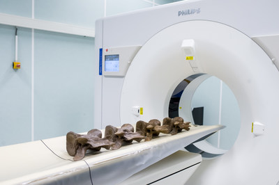
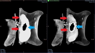
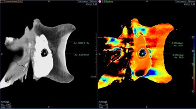
AMSTERDAM, Sept. 28, 2016 /PRNewswire/ -- Royal Philips (NYSE: PHG, AEX: PHIA), a leading health technology company, today presented the results of a Spectral CT scan of the tail vertebrae of Trix, a 66 million year old Tyrannosaurus Rex dinosaur. Working with Leiden's Naturalis museum in the Netherlands, Philips offered to scan one of the world's most complete and best preserved T. Rex skeletons. The aim of the scan was to evaluate how images of Trix's tail vertebrae obtained using Philips' IQon Spectral CT imaging differ from conventional CT images, and to provide a level of detail not visible to paleontologists until now.

Trix, who was discovered in Montana, USA, in 2013, was purchased by the Naturalis museum and transported to the Netherlands on August 23, 2016. For 66 million years this carnivorous dinosaur lay buried in the rocks. As the organic matter had virtually all disappeared, only the skeleton was left, and over the millennia a mineralization process took place. As a result, Trix's fossilized bones can be considered 'remineralized' representations of her original bones.
The smaller bones in the tail of a T. Rex fossil are often missing, lost to scavenging or washed away by currents prior to burial and fossilization. Intact smaller tail elements of a T. Rex are therefore rare, and Philips was not aware of much prior work on the internal structure of these elements. However, Trix has a beautifully preserved tail. Paleontologist and dinosaur expert Anne Schulp of Naturalis was keen to examine Trix's vertebrae in greater detail. On August 31, her four tail vertebrae (C20 C23) were scanned on a Philips IQon Spectral CT scanner in Best, in the Netherlands.
"We have already looked at some of the bones using a regular medical CT scanner, but we had a little problem," said Anne Schulp, Paleontologist and dinosaur expert at Naturalis. "There is a deposit of pyrite an iron rich mineral that blurs the complete picture because it absorbs the X-rays. So basically we required a different technology."

Philips IQon is the world's first detector-based spectral CT scanner. In addition to its unique dual-layer detector, it makes use of advanced spectral reconstruction algorithms. By combining these two technologies, the IQon is able to obtain detailed images of objects that are otherwise difficult to scan, such as Trix's tail vertebrae.
"The IQon filters out all the 'noise,' as it were, thereby providing a really good insight into the bone structure and how it is built up," explained Anne Schulp. "It's basically making the step from black and white movies to color. In this way, we are able to add depth to Trix's medical records: we make the invisible visible. It's just really hard to describe the sensation of finding something, seeing something that no one else has ever seen."
These scans are just the first images. Further investigations will provide even more insights to build up Trix's medical record. Over the next few months, on-going research with medical experts will aim to reveal more details about Trix's life.

"What the IQon is doing is acquiring two different X-ray energy levels in a single acquisition. It's fantastic to see how a detailed three-dimensional image is built up," said Julien Milles, Clinical Scientist CT Health Systems, Philips Benelux. "With Trix, we were able to identify and color-highlight bone structure, even with 'remineralized' versions of the original bones. So in one picture it is just one white blob. And then all of a sudden, at the click of a mouse, there is the next one where all the details in the bone pop up. And that with a 66 million year old lady!"
This isn't the first time Philips' imaging technology has been leveraged for unearthing secrets of the past. In 2015, Philips' CT scanners were used to study bones of the bodies of people who died in Pompeii, giving archeologists a view into the daily lives, health and habits of the citizens of Pompeii.
For more information on the Philips IQon Spectral CT, visit www.philips.com/iqon.
For further information, please contact:
Marianne Nouwens
Philips Benelux
Tel.: +31 6 1158 1004
E-mail: Marianne.Nouwens@Philips.com
Kathy O'Reilly
Philips Group Communications
Tel.: +1 978-229-8919
E-mail: Kathy.oreilly@philips.com
About Royal Philips
Royal Philips (NYSE: PHG, AEX: PHIA) is a leading health technology company focused on improving people's health and enabling better outcomes across the health continuum from healthy living and prevention, to diagnosis, treatment and home care. Philips leverages advanced technology and deep clinical and consumer insights to deliver integrated solutions. Headquartered in the Netherlands, the company is a leader in diagnostic imaging, image-guided therapy, patient monitoring and health informatics, as well as in consumer health and home care. Philips' health technology portfolio generated 2015 sales of EUR 16.8 billion and employs approximately 69,000 employees with sales and services in more than 100 countries. News about Philips can be found at www.philips.com/newscenter
Photo - http://photos.prnewswire.com/prnh/20160928/412788
Photo - http://photos.prnewswire.com/prnh/20160928/412789
Photo - http://photos.prnewswire.com/prnh/20160928/412828
To view the original version on PR Newswire, visit:http://www.prnewswire.com/news-releases/philips-colors-t-rex-trixs-tail-using-its-spectral-ct-technology-300335677.html
SOURCE Royal Philips


