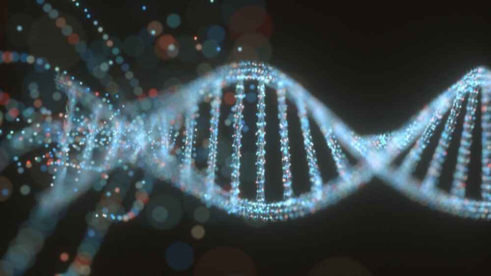
A new technique, DNA microscopy, is allowing scientists to visualize DNA in a way not previously possible. And there isn’t even an actual microscope involved.
When scientists need to see something too small for a microscope, what do they do? A new technique, DNA microscopy, is allowing scientists to visualize DNA in a way not previously possible. And there isn’t even an actual microscope involved.
Scientific experiments rely on many different types of data in order to form and test questions about biology. Some of those experiments provide indirect evidence about what is occurring, however, microscopes have long been used to see things we cannot view with the naked eye. The first microscopes used light and glass to magnify what was living in the water. For centuries, this let scientists “see it and believe it.”
The familiar silhouette of the microscope has become a staple of scientific exploration. However, every tool has its limits, and the microscope is no exception.
When it comes to the microscope, limitations come in the form of size and resolution. Size speaks to how small a user can see. Some microscopes can illuminate the details of fibers, while others are strong enough to discern proteins within cells.
There are many types of microscopes. For example, instead of light, some microscopes use electrons to peek inside of the microscopic world. A microscope’s resolution is how well you can distinguish between the tiny things you are looking at. A fuzzy image of an object taken with a higher resolution microscope may reveal that you were actually looking at two separate objects that were too close together to be distinguished.
So what happens when something goes below those limits, making something difficult to see clearly? That has long been a concern for visualizing DNA, the code of life that exists in each of our cells. Current techniques to visualize DNA involve making it fluoresce against a background, typically using a dye known as DAPI.
While DAPI staining identifies where DNA is, it doesn’t give genomic information. Pairing genomic information with location information for cells is critical to understanding systems. For example, many tumorous cancers are heterogeneous, meaning that the mutations you find in one part of the tumor may be very different than what you find in another part of the tumor. Understanding where those mutations occur and how could help researchers understand how tumors form and possibly how to better treat them.
A new molecular technique, DNA microscopy, more easily obtains both sequence and spatial information about DNA regions of interest.
The process works by tagging the DNA (or the workhorse copies of DNA,called RNA) with barcodes. These barcodes work similarly to barcodes on products you buy from the store. When researchers add them to fixed cells, the barcodes attach to particular “products”, in this case, RNA molecules. At the end of the process, the barcodes can help identify each molecule, just like the register at the store can scan the barcode and know which brand of cereal you are buying. However, unlike barcodes on products, these barcodes are just as small as the products they are tagging.
To amplify the signal that the barcodes can broadcast, copies are created of the tagged molecules. As more and more copies are made, the barcodes begin to collide. They stick together and become linked. This process of amplifying, colliding and linking continues, expanding the signal of the barcode out from the molecules’ original location.
Perhaps the most impressive part of this microscopy technique is that there’s actually no microscope involved. Instead, the now-linked barcodes are sequenced, revealing which barcodes are stuck to each other and how often.
The underlying principle is that molecules close to each other will collide far more often than molecules that are further apart. This information can then be reconstructed with an algorithm developed by the researchers to create a picture of how the regions are spatially arranged within the cell and how cells are spatially arranged compared to each other. Scientists are able to reconstruct a 3-dimensional image based on how the barcode signals interacted as the amplification process happened.
Researchers can now learn how DNA organization connects to its genomic context in different conditions. Allowing scientists to connect genomic location and spatial information opens a new world of questions that can be addressed.


